

 See My Latest Article on HelloNation
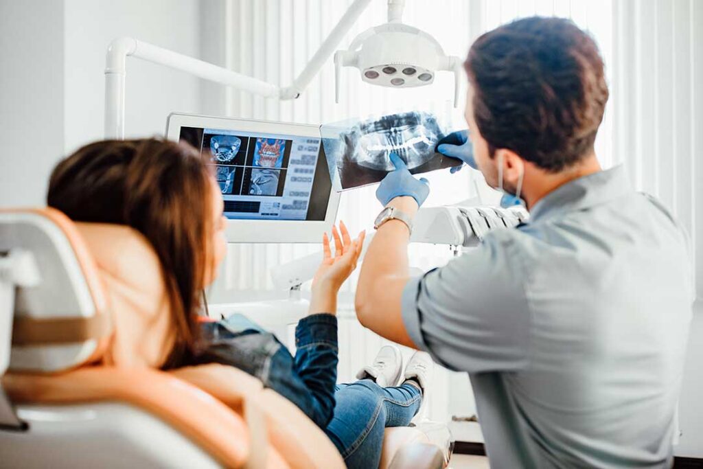
Dental X-rays are diagnostic images that help reveal problems a visual examination cannot always detect. They can show decay between teeth, changes in jawbone, developing teeth, infection, and other conditions before symptoms become obvious.
For residents of Moses Lake, understanding why an X-ray is recommended can make routine dental care feel less uncertain. The type and timing of imaging should be based on a person’s age, oral health history, symptoms, and risk factors—not on a fixed schedule for everyone.
Why are dental X-rays needed?
Dental X-rays help examine areas hidden from view, including the spaces between teeth, tooth roots, jawbone, and developing permanent teeth. A person may have no pain even when a cavity, bone change, or infection is present.
X-rays may help identify:
- Cavities between teeth or beneath existing fillings
- Bone loss associated with gum disease
- Abscesses or infections near tooth roots
- Cracks, impacted teeth, or unusual tooth development
- Cysts, tumors, or other changes in the jaw
- The position of permanent teeth in children
- Dental injuries that affect roots or surrounding bone
An X-ray is not a replacement for an examination. Instead, it adds information that cannot be obtained by looking at the teeth and gums alone.
How often should dental X-rays be taken?
There is no universal schedule that applies to every patient. The appropriate interval depends on oral health, age, previous findings, symptoms, tooth decay risk, gum health, and whether a person has recently changed providers or had incomplete records transferred.
Someone with a history of frequent cavities may need imaging more often than someone with stable oral health and a low risk of decay. Children and teenagers may also need different imaging because their teeth and jaws are still developing.
New X-rays may be reasonable when:
- A new patient has no recent images available
- A toothache, swelling, sensitivity, or injury needs evaluation
- Earlier images are too old or do not include the area of concern
- Treatment is being planned
- A change in oral health suggests that earlier images may no longer be sufficient
Routine repetition without a clinical reason is generally avoided. Previous X-rays can sometimes be transferred between dental offices, reducing the need to take duplicate images.
What are the main types of dental X-rays?
Different images answer different questions. A small image of one area is not interchangeable with a broad view of the jaws.
Bitewing X-rays show the upper and lower back teeth in the same image. They are commonly used to detect cavities between teeth and to evaluate bone levels.
Periapical X-rays capture an entire tooth, including the root and the surrounding bone. They may be used to investigate pain, infection, trauma, or changes near the root.
Panoramic X-rays provide a broad view of the upper and lower jaws, teeth, jaw joints, and surrounding structures. They may be useful for evaluating wisdom teeth, orthodontic development, jaw injuries, or certain growth patterns.
Cephalometric X-rays are side-view images often used when evaluating jaw relationships or planning orthodontic treatment.
Cone-beam computed tomography, sometimes called CBCT, creates three-dimensional images. It provides more detail than ordinary dental X-rays but also involves more radiation than many two-dimensional images. Because of that, it should be used when the additional information is likely to affect diagnosis or treatment.
Are dental X-rays safe?
Dental X-rays use a low amount of ionizing radiation, and modern equipment is designed to limit exposure. The goal is to obtain a useful image with the lowest reasonable dose.
Radiation safety is often described by the principle “as low as reasonably achievable.” In practice, this means the image should have a clear medical or dental purpose, the area being examined should be limited when possible, and unnecessary repeat images should be avoided.
A patient can reasonably ask:
- What area will this X-ray examine?
- What question is the image intended to answer?
- Are previous images available?
- Would waiting or using another type of image change the decision?
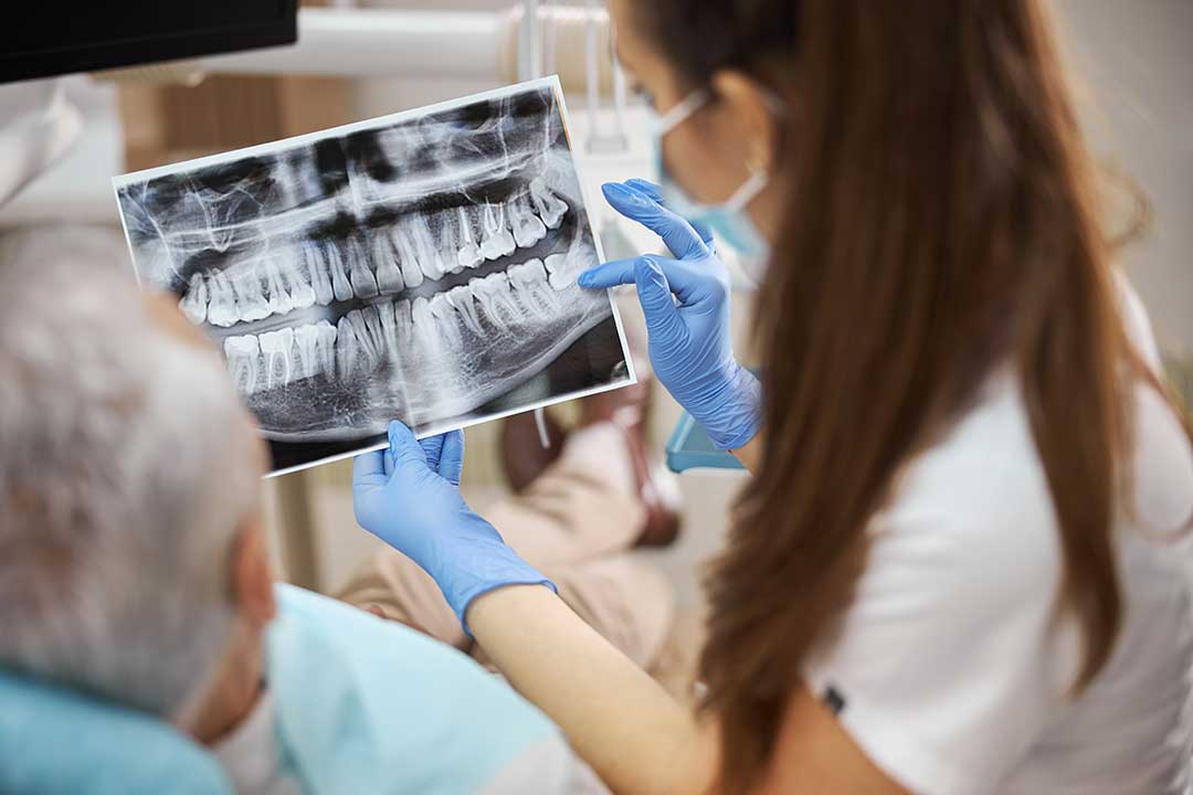
These questions are part of informed care and do not suggest that a patient is refusing necessary treatment.
What if someone is pregnant or might be pregnant?
Tell the dental team about a known or possible pregnancy before imaging. Dental X-rays may still be appropriate during pregnancy when they are needed to evaluate pain, infection, injury, or another dental problem.
The amount of radiation from dental imaging is directed toward the mouth and is very small. Delaying necessary care can allow an infection or other condition to worsen. For nonurgent imaging, the timing may be discussed based on the individual situation.
Pregnancy should not be treated as a reason to ignore severe tooth pain, facial swelling, fever, or other warning signs. These symptoms may require timely evaluation.
Are dental X-rays safe for children?
Children are more sensitive to radiation than adults, and their teeth and jaws are still changing. That does not mean X-rays should be avoided altogether. It means imaging should be selected carefully and performed only when it can provide useful information.
Children may need X-rays to check for:
- Cavities that cannot be seen between baby teeth
- The position and development of permanent teeth
- Delayed eruption or unusual tooth placement
- Injuries to developing teeth
- Jaw growth or orthodontic concerns
The number and type of images should match the child’s age, dental history, symptoms, and risk of disease. A child does not automatically need a full set of X-rays at every visit.
What happens during the procedure?
Most dental X-rays take only a few minutes. A small sensor or image receptor is placed in the mouth, and the patient is asked to remain still while the image is captured. Some people find the sensor uncomfortable, especially if the mouth is tender or the gag reflex is strong.
Panoramic imaging usually involves standing or sitting still while the equipment rotates around the head. The process is painless, although positioning may feel unfamiliar.
If the sensor causes discomfort, tell the dental team. Small adjustments in positioning, breathing, or timing may make the procedure easier.
What should Moses Lake residents consider?
Dry conditions, seasonal routines, and busy household schedules can make it easy to postpone care until a problem becomes painful. In a community where some residents may travel a significant distance for appointments or coordinate care around work, school, and family responsibilities, keeping copies of recent dental images can be practical.
People who move between dental offices, change insurance arrangements, or receive urgent care away from home may benefit from knowing when their last X-rays were taken and what type they were. This information can help prevent unnecessary duplication while ensuring that important areas are not overlooked.
When should a dental problem be evaluated promptly?
An X-ray may be especially useful when symptoms suggest a problem beneath the surface. Prompt evaluation is appropriate for facial swelling, fever with dental pain, a broken tooth, persistent sensitivity, pain when biting, unexplained bleeding, or an injury to the mouth or jaw.
Severe swelling that affects breathing or swallowing requires emergency medical attention.
A dental X-ray is a focused diagnostic tool, not a routine exposure that must be repeated automatically. The most useful approach is individualized: consider the patient’s symptoms and history, use the smallest appropriate examination, and avoid imaging that is unlikely to change care.